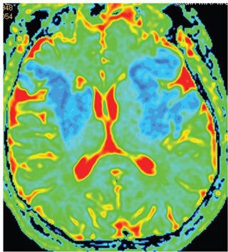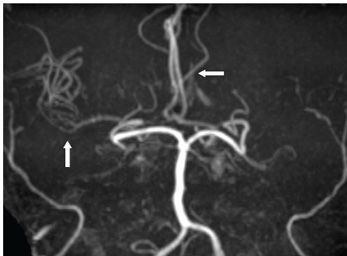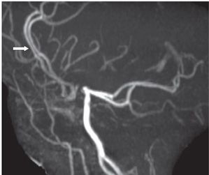


FINDINGS These images were obtained on the second day of admission. Day 1 images (not shown) showed a left perisylvian area of restricted diffusion. Figures 286-1 and 286-2. Axial DWI and corresponding ADC map through the lateral ventricles. There is bilateral perisylvian restricted diffusion. The right basal ganglia is involved, while the left posterior lentiform nucleus and the adjacent left corona radiata are involved as well. Inferior extension into the anterior temporal lobes (not shown) was present bilaterally. Figures 286-3 and 286-4. 3D time of flight MRA of the head obtained on day 1 showing nonvisualization of bilateral internal carotid arteries and the left middle cerebral artery (MCA). The right MCA (vertical arrow) is attenuated. Its occlusion is probably responsible for the new infarct on the right side. Bilateral anterior cerebral arteries (ACAs) (transverse arrows) show robust intensity.
DIFFERENTIAL DIAGNOSIS N/A.
DIAGNOSIS Bilateral MCA territory acute infarction.
DISCUSSION
Stay updated, free articles. Join our Telegram channel

Full access? Get Clinical Tree


