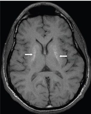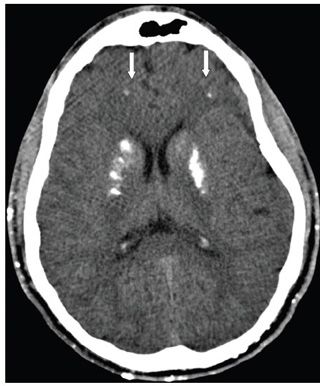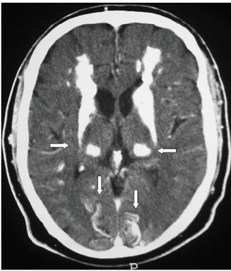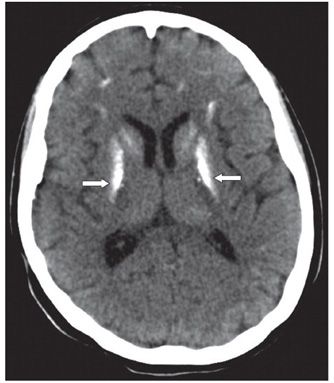


FINDINGS Figure 287-1. Axial NCCT through the basal ganglia. There are bilateral symmetrical calcifications in the basal ganglia, the lentiform nuclei and caudate heads (transverse arrows). There is a small punctate calcification in the white matter (WM) of the right frontal lobe (vertical arrow). Figure 287-2. Corresponding non-contrast MR T1WI through the basal ganglia. The calcifications are slightly hyperintense (transverse arrows). Figure 287-3. Axial NCCT in a patient with renal failure and secondary parathyroid disease. There is calcifications in the basal ganglia and frontal WM (vertical arrows) and also superficially in scalp. Figure 287-4. In a patient with Fahr disease this axial NCCT shows extensive calcifications in the deep gray matter (GM), basal ganglia and thalami (transverse arrows), WM, and occipital cortex (vertical arrows). Figure 287-5
Stay updated, free articles. Join our Telegram channel

Full access? Get Clinical Tree


