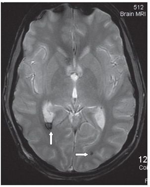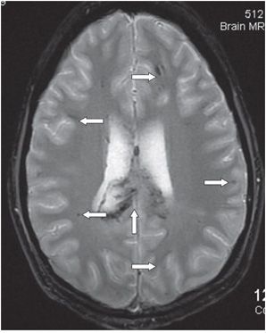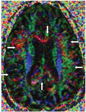


FINDINGS Figure 288-1. Axial MRI GRE through the fourth ventricle. There are multifocal bilateral cerebellar, pons, and left superior cerebellar peduncle punctate hypointensities (arrows show some of them) consistent with hemorrhagic products. Figure 288-2. Axial GRE through the trigones. There is hypointense sedimentation with fluid-fluid level in the right trigone consistent with intraventricular hemorrhage (vertical arrow). There is visible subcortical punctate hypointensity in the left occipital lobe (transverse arrow). Figure 288-3. Axial GRE through the corpus callosum (CC). There is heterogeneous enlargement of the posterior body and splenium of the CC (vertical arrow). There are numerous bilateral frontal and parietal subcortical hypointensities (transverse arrows identify some of them). Figure 288-4. Color directional MRI DTI map through the CC. There is disorganization and asymmetry of fiber tracts (transverse arrows). There is disruption, enlargement, and heterogeneity of the usual red transverse fiber tracts of the CC (vertical arrows).
DIFFERENTIAL DIAGNOSIS N/A.
DIAGNOSIS Traumatic brain injury with diffuse axonal injury (TBI/DAI).
DISCUSSION
Stay updated, free articles. Join our Telegram channel

Full access? Get Clinical Tree


