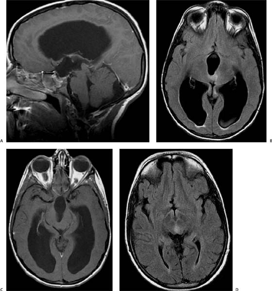
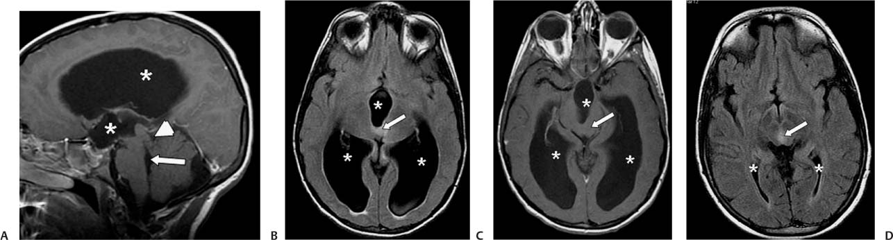
Case 29 A 12-year-old boy with progressive headaches. (A) Sagittal T1-weighted image (WI) shows noncommunicating hydrocephalus with dilated lateral and 3rd ventricles (asterisks). The 4th ventricle is not dilated (arrow), and the anatomy of the tectum is distorted (arrowhead). (B) Axial fluid-attenuated inversion recovery (FLAIR) image shows hyperintensity in the midbrain (arrow). The lateral and 3rd ventricles are dilated (asterisks). (C) Axial T1WI with contrast shows no enhancement in the midbrain (arrow). Hydrocephalus is seen (asterisks). (D) Axial FLAIR image shows normal size of the lateral ventricles (asterisks) after decompression through a shunt. The area of hyperintensity in the midbrain is again seen (arrow).
Clinical Presentation
Imaging Findings
Differential Diagnosis
Stay updated, free articles. Join our Telegram channel

Full access? Get Clinical Tree


