 Clinical Presentation
A 74-year-old man with right-sided chest pain and weight loss.
 Imaging Findings
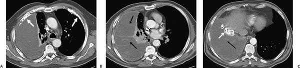
(A–C) Contrast-enhanced computed tomography of the chest shows a large right-sided pleural opacity with ill-defined linear areas of enhancement (black arrows), as well as densely calcified pleural plaques bilaterally, including a calcified plaque on the right diaphragmatic surface (white arrows).
 Differential Diagnosis
• Malignant pleural mesothelioma (MPM): Enhancing tissue and calcified pleural plaques are suggestive of asbestos related pleural mesothelioma.
• Loculated empyema:
Stay updated, free articles. Join our Telegram channel

Full access? Get Clinical Tree


