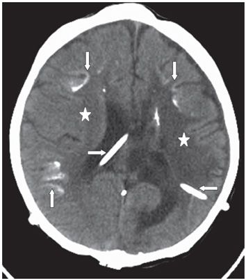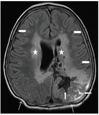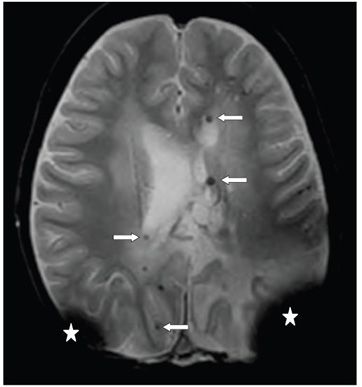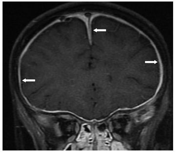


FINDINGS Figure 29-1. Axial treatment planning post-contrast T1WI through the lateral ventricles. There is a large partially cystic and solid mass (WHO IV) astrocytoma (star) of the corpus callosum bulging into the lateral ventricles. Figure 29-2. Axial NCCT through the corona radiata about 2 years into his treatment which included surgical removal of the tumor via bilateral parietal craniotomies, radiation treatment, and chemotherapy. There are bilateral ventriculoperitoneal (VP) shunts (transverse arrows), multifocal subcortical calcifications (vertical arrows), and bilateral periventricular smudgy white matter (WM) hypodensities (stars). Figure 29-3. Axial FLAIR through the corona radiata. There is bilateral confluent WM hyperintensity (stars). The left parasagittal parietal surgical cavity (vertical arrow) represents part of the surgical tract to the original tumor. There are hyperintensities in the left parietal sulci (transverse arrow) produced by the VP shunt susceptibility artifact. There is thickening of the dura (chevrons). The craniotomy flap is present posteriorly (line arrows). Figure 29-4. Axial GRE through the centrum semiovale. There are bilateral large signal void artifacts from the VP shunts (stars). There are multiple punctate hypointensities in bilateral centrum semiovale WM (transverse arrows) consistent with telangiectasia or microhemorrhages or calcifications. Figure 29-5
Stay updated, free articles. Join our Telegram channel

Full access? Get Clinical Tree


