CASE 29
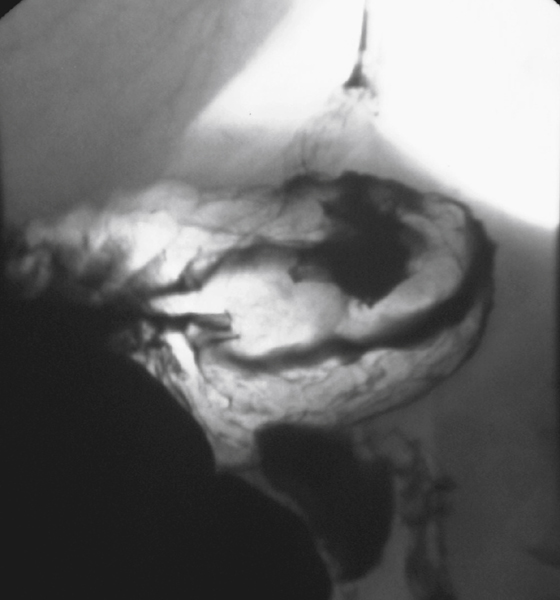
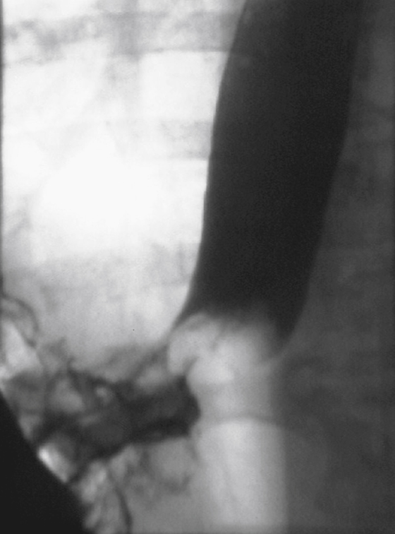
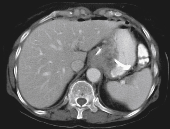
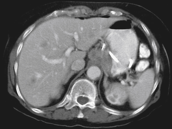
History: A 44-year-old man presents with dysphagia.
1. Which of the following should be included in the likely differential diagnosis of the imaging findings? (Choose all that apply.)
B. Esophageal squamous cell carcinoma
C. Primary esophageal lymphoma
E. Gastric squamous cell carcinoma
2. What is the most common type of malignancy seen at the gastroesophageal junction?
3. There are non-neoplastic processes that could give this appearance of a mass at the gastro-esophageal junction. Which of the following would not be expected to give this appearance?
Stay updated, free articles. Join our Telegram channel

Full access? Get Clinical Tree


