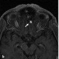
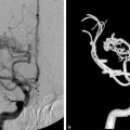
Fig. 29.1 Selective sphenopalatine injection in lateral (a) and anteroposterior (AP) (b) views demonstrates multiple telangiectasias of the nasal mucosa, as classically seen in HHT. They are fed by the medial (septal) and lateral (conchal) arteries of the distal sphenopalatine artery.
29.1.3 Diagnosis
Nosebleeds resulting from multiple mucocutaneous telangiectasias of the distal sphenopalatine arteries, suggestive of hereditary hemorrhagic telangiectasia (HHT).
29.2 Anatomy
The vascular supply to the nasal fossa involves both external and internal carotid arterial contributions, with four major contributing vessels involved on each site.
First, the sphenopalatine artery is the major contributor for the posteromedial and posterolateral supply to the nasal fossa. This vessel is the most important source of supply to the mucoperiosteum of the nose. It originates from the pterygopalatine segment of the maxillary artery. The sphenopalatine artery exits from the superomedial aspect of the pterygopalatine fossa via the sphenopalatine foramen and enters the nasal fossa behind and slightly above the middle concha. It has two major groups of branches: posterior lateral nasal and posterior medial (or septal) branches. The posterior lateral nasal arteries supply the conchae and can anastomose with the anterior and posterior ethmoidal arteries superiorly. After the posterior lateral nasal artery has branched off the sphenopalatine artery, its main trunk courses medially along the posterior roof of the nasal cavity. Once the vessel has reached the septum, it gives origin to the posterior medial (or septal) branches that run anteriorly along the midline. The inferior septal branch courses as the nasopalatine artery through the incisive canal to anastomose with the greater palatine artery. Superiorly, small branches run toward the cribriform plate and anastomose with nasal branches of the ethmoidal arteries (▶ Fig. 29.2).
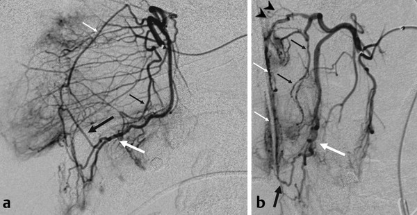
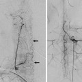
Fig. 29.2 Anatomy of the internal maxillary artery branches supplying the nose in lateral (a) and AP (b) views. The sphenopalatine artery has medial septal (small white arrows) and lateral conchal (small black arrows) branches. At the midline, the superior medial branches anastomose through the cribriform plate with the ethmoidal branches (arrowheads) of the ophthalmic artery. The inferior medial branch anastomoses with the greater palatine artery (large white arrow) as it courses as the nasopalatine artery (large black arrow) through the incisive canal.
Second, the anterior and posterior ethmoidal arteries supply the superior aspect of the nasal fossa. Both arteries arise from the ophthalmic artery and send numerous small branches through the cribriform plate that anastomose medially and laterally with nasal branches of the sphenopalatine artery and provide a potential collateral pathway between the internal and external carotid circulations, which may be a cause for failed control of epistaxis after external carotid artery (ECA) embolization.
Third, the terminal branch of the greater palatine artery supplies the inferior medial and lateral part of the nasal cavity. It enters the incisive foramen and anastomoses with the nasopalatine artery (i.e., the inferior septal branch of the sphenopalatine artery).
Finally, the septal branch of the superior labial artery that originates from the facial artery supplies the medial inferior wall of the nasal cavity. At the anteroinferior medial wall, the superior labial artery anastomoses with both the sphenopalatine artery (via the nasopalatine artery) and the distal greater palatine artery. This area is also known as locus Kiesselbachii and is the most common culprit region for anterior epistaxis.
29.3 Clinical Impact
29.3.1
Stay updated, free articles. Join our Telegram channel

Full access? Get Clinical Tree


