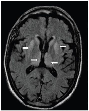
FINDINGS Figure 291-1. Axial T2WI through the thalami. There is subtle hyperintensity in the basal ganglia and medial and posterior thalami bilaterally and symmetrically. Figure 291-2. The abnormalities are more obvious and better seen on the corresponding FLAIR image (arrows).
DIFFERENTIAL DIAGNOSIS: IMAGING Postinfectious encephalitis, Creutzfeldt-Jacob disease (vCJD), cat-scratch disease, intracranial hypertension, and Alpers syndrome.
DIAGNOSIS Variant form of Creutzfeldt-Jacob disease (vCJD).
DISCUSSION


Stay updated, free articles. Join our Telegram channel

Full access? Get Clinical Tree


