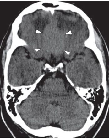
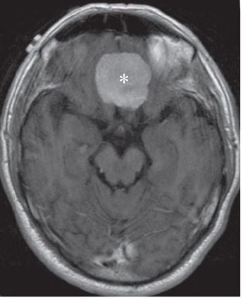
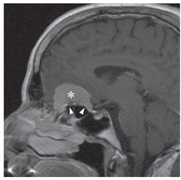
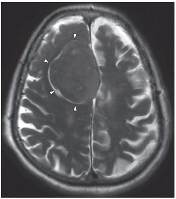
FINDINGS Figures 292-1 and 292-2. Contiguous NCCT through inferior frontal lobes. There is bilateral frontal lobe edema (arrows in Figure 292-1) and mild mass effect (partial regional sulcal effacement) secondary to an isodense midline mass along the anterior skull base (arrowheads in Figure 292-2). Figures 292-3 and 292-4. Axial and coronal post-contrast T1WI demonstrates homogeneous avid enhancement of the dural-based, extraaxial mass (asterisk). This tumor covers the skull base from the posterior margin of the olfactory groove through the planum sphenoidale, nearly reaching the tuberculum sella. Note the upward doming, or “blistering” of the sphenoid sinus roof toward the tumor on the sagittal image, a feature known as pneumosinus dilatans (arrowheads in Figure 292-4). The cribriform plate was not transgressed by the tumor in this case. Figure 292-5. Axial T2WI in a comparison case of a right frontal parafalcine meningioma. The mass is isointense with gray matter (GM). There is a cerebrospinal fluid (CSF) cleft between the mass and the brain parenchyma (arrow heads) defining the extraaxial location of the mass.
DIFFERENTIAL DIAGNOSIS Dural metastasis, hemangiopericytoma, meningioma.
DIAGNOSIS Meningioma.
DISCUSSION


Stay updated, free articles. Join our Telegram channel

Full access? Get Clinical Tree


