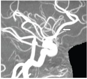
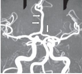
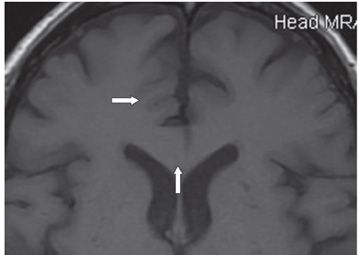
FINDINGS Figure 293-1. Axial source image from 3D TOF MRA of the head through the third ventricle. There is a single anterior cerebral artery (ACA) within the anterior interhemispheric fissure (arrow). Figure 293-2. Lateral MIP 3D MRA of the brain. There is a single ACA A2 (arrow). Figure 293-3. Submentovertical 3D TOF MRA of the brain. There are two A1s joining together to form a single A2 (transverse arrows). There is no anterior communicating artery. The left A1 is mildly hypoplastic (vertical arrow). Figure 293-4. Axial non-contrast T1WI. Asymmetric frontal medial sulcation abnormality; right parasagittal microgyria (arrow). There is mildly malformed thickening of the genu of corpus callosum (vertical arrow).


Stay updated, free articles. Join our Telegram channel

Full access? Get Clinical Tree


