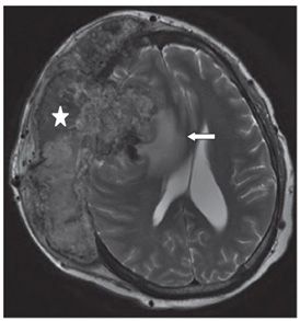
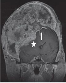
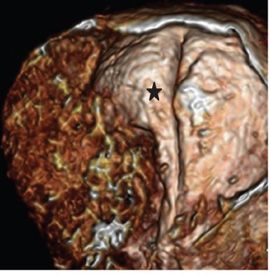
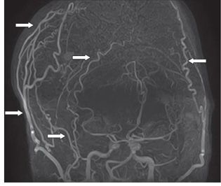
FINDINGS Figure 294-1. Axial NCCT bone window through the cranial vault superiorly. There is a rather irregular knobbly 16.4 cm × 8.1 cm right hemicranial bony mass involving the right frontotemporoparietal bones with patchy lytic and sclerotic areas projecting both intracranially and extracranially (arrows). Figure 294-2. Axial T2WI through the body of the lateral ventricles. The mass is heterogeneous and of mixed intensity (star). There is a large component projecting intracranial compressing the right frontal lobe, right lateral ventricle and displacing midline structures (falx) to the left. There is a right frontal white matter (WM) hyperintensity adjacent to the intracranially projecting mass consistent with vasogenic edema (arrow). Figure 294-3


Stay updated, free articles. Join our Telegram channel

Full access? Get Clinical Tree


