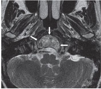
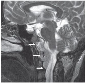
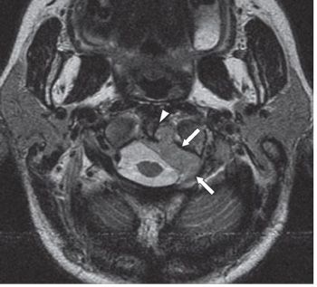
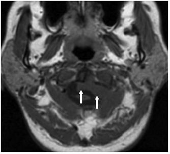
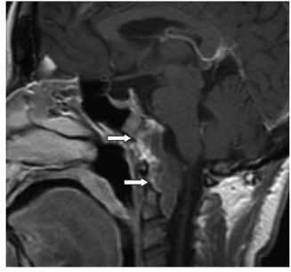
FINDINGS Figure 295-1. Axial NCCT at the level of foramen magnum. There is a large, midline, rounded region of geographic bony remodeling in the basion/clivus with a narrow, nonsclerotic zone of transition (arrows). Note the absence of calcification in the lesion. Figures 295-2 and 295-3. Axial, small FOV T2WI and sagittal T2WI MRI at the same level, respectively. There is a corresponding mass of mixed/heterogeneous intensity (arrows) anterior to brainstem and spinal cord. Figure 295-4. A slightly more inferior axial T2WI (at the level of the anterior C1–C2 joint). There is tumor extension into the ventral and left lateral aspects of the spinal canal (arrows) eroding the dens (arrowhead). Figure 295-5. The corresponding axial T1WI. The mass (arrow) is isointense to surrounding muscle. Figure 295-6. Sagittal post-contrast T1WI. The mass enhances heterogeneously primarily along the ventral margin of the tumor (arrow).
DIFFERENTIAL DIAGNOSIS Metastasis, chordoma, chondrosarcoma, plasmacytoma, extralymphatic/high-grade non-Hodgkin lymphoma, meningioma.
DIAGNOSIS Chordoma of the clivus.
DISCUSSION


Stay updated, free articles. Join our Telegram channel

Full access? Get Clinical Tree


