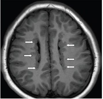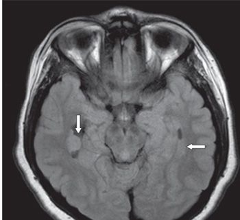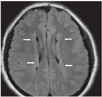


FINDINGS Figure 296-1. Axial T1WI through the temporal horns. There is a subependymal nodule isointense with cortical gray matter (GM) projecting into the right temporal horn (vertical arrow). Similar but less defined nodules surround the left temporal horn (transverse arrow). Figure 296-2. Axial T1WI through the body of the lateral ventricles. There are multiple bilateral symmetrical contiguous subependymal nodules isointense with cortical GM projecting into the lateral ventricles (arrows). Figures 296-3 and 296-4. Axial FLAIR through the temporal horns and body of the lateral ventricles, respectively. There are multiple symmetrical isointense (to GM) contiguous subependymal nodules projecting into the temporal horns and body of the lateral ventricles (arrows).
DIFFERENTIAL DIAGNOSIS Heterotopic GM, tuberous sclerosis complex, toxoplasmosis, subependymoma.
DIAGNOSIS Periventricular nodular GM heterotopia (PNH).
DISCUSSION
Stay updated, free articles. Join our Telegram channel

Full access? Get Clinical Tree


