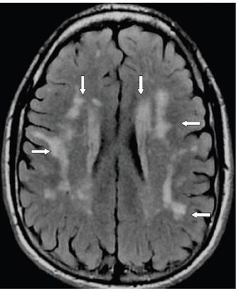
FINDINGS Figures 297-1 and 297-2. Axial FLAIR through the temporal lobes and the corona radiata, respectively. There are multiple hyperintense foci throughout the subcortical, deep, and periventricular WM of the temporal lobes in Figure 297-1 and bilateral corona radiata in Figure 297-2 (arrows). The lesions did not enhance nor restrict diffusion (images are not provided).
DIFFERENTIAL DIAGNOSIS Multiple sclerosis (MS), sporadic subcortical arteriosclerotic encephalopathy (sSAE), primary angiitis of central nervous system (CNS), posterior reversible encephalopathy syndrome (PRES), mitochondrial encephalopathy with lactic acidosis and strokes (MELAS), cerebral autosomal dominant arteriopathy with subcortical infarcts and leukoencephalopathy (CADASIL).
DIAGNOSIS CADASIL.
DISCUSSION
Stay updated, free articles. Join our Telegram channel

Full access? Get Clinical Tree


