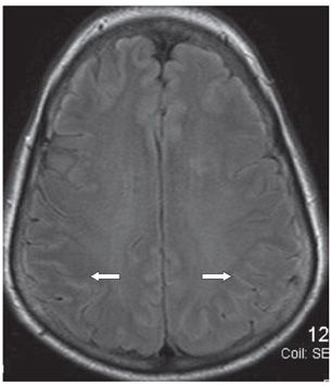
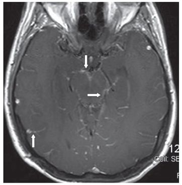
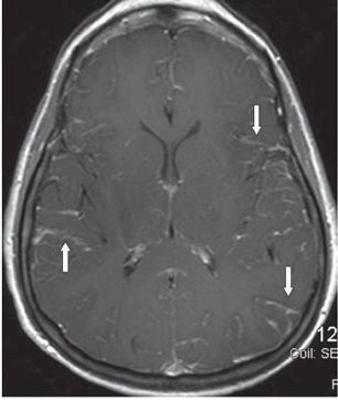
FINDINGS Figures 298-1 and 298-2. Axial FLAIR through the midbrain and the centrum semiovale. There is widespread sulcal hyperintensity (arrows). Figures 298-3 and 298-4. Axial post-contrast T1WI through the midbrain and the sylvian fissures in the same patient. There is extensive mostly linear subarachnoid space enhancement (arrows) around the midbrain in Figure 298-3 and in the sylvian fissures and convexity sulci in Figure 298-4.
DIFFERENTIAL DIAGNOSIS Leptomeningeal enhancement (LME) due to meningitis, vasculitis, carcinomatosis, lymphoma.
DIAGNOSIS LME due to migraine headache.
DISCUSSION


Stay updated, free articles. Join our Telegram channel

Full access? Get Clinical Tree


