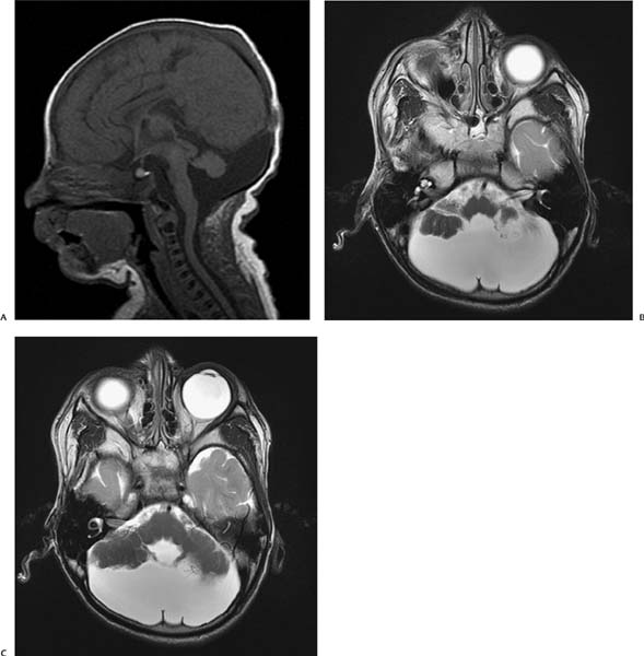
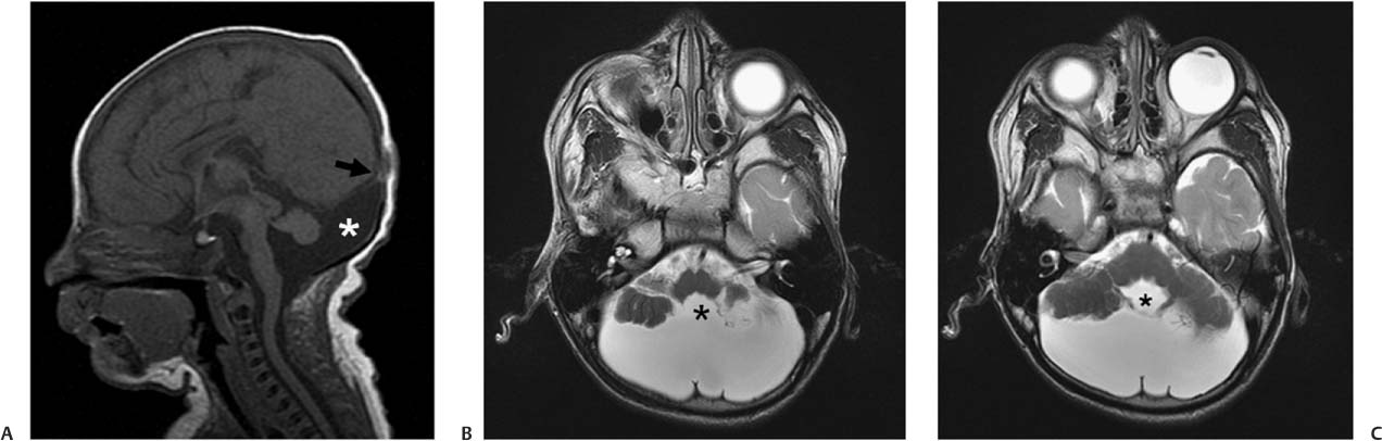
Case 3 A 4-year-old boy with developmental delay and seizures. (A) Sagittal T1-weighted image (WI) of the brain demonstrates a large posterior fossa and an elevated insertion of the tentorium (arrow) with respect to the expected position. A cyst (asterisk) that communicates with the 4th ventricle fills the posterior fossa. (B,C) Axial T2WIs of the brain show cystic dilatation of the 4th ventricle (asterisks), which is ballooning between hypoplastic cerebellar hemispheres. The cerebellar vermis is absent. • Dandy-Walker continuum: The high insertion of the tentorium and cystic dilatation of the 4th ventricle are typical features of Dandy-Walker continuum. The degree of cerebellar hypogenesis may vary. • Mega cisterna magna:
Clinical Presentation
Imaging Findings
Differential Diagnosis
![]()
Stay updated, free articles. Join our Telegram channel

Full access? Get Clinical Tree


