 Clinical Presentation
A 55-year-old woman with a history of epistaxis and headache.
 Imaging Findings
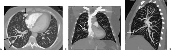
(A–C) Contrast-enhanced computed tomography (CT) of the chest. Axial (A), coronal (B), and sagittal (C) maximum-intensity-projection reconstructions demonstrate multiple dilated vascular structures with prominent connecting vessels throughout the right lung, consistent with arteriovenous (AV) malformations (arrows).
 Differential Diagnosis
• Hereditary hemorrhagic telangiectasia (HHT; Osler-Weber-Rendu disease):
Stay updated, free articles. Join our Telegram channel

Full access? Get Clinical Tree


