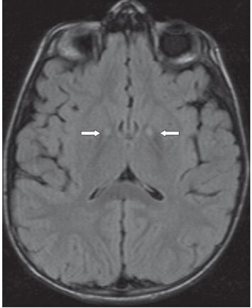
FINDINGS Figure 3-1. Axial T2WI through the basal ganglia. There is bilateral focal symmetrical hyperintensity in the medial globus pallidi (due to gliosis and spongiosis) surrounded by thin hypointensity (due to iron and other metal deposition) (arrows) compatible with the “eye of the tiger” sign. Figure 3-2. Corresponding FLAIR image shows similar but perhaps less obvious findings.
DIFFERENTIAL DIAGNOSIS Multiple system atrophy, cortical basal ganglionic degeneration, multiple sclerosis, neurofibromatosis, human immunodeficiency virus dementia, Freidrich ataxia, carbon monoxide poisoning, PKAN.
DIAGNOSIS Pantothenate kinase-associated neurodegeneration (PKAN).
DISCUSSION
Stay updated, free articles. Join our Telegram channel

Full access? Get Clinical Tree


