Case 3
 Clinical Presentation
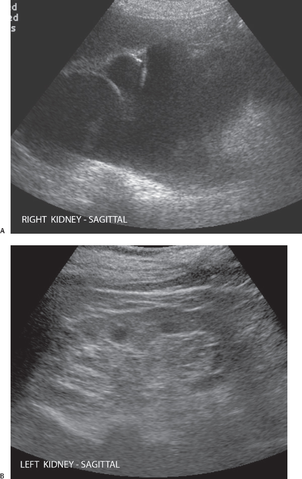
A 74-year-old woman with a history of fullness in the right flank.
 Imaging Findings
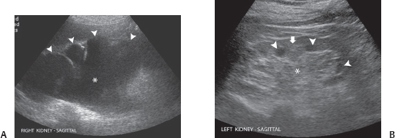
(A) Longitudinal sonographic image of the right kidney shows complete atrophy of the right renal parenchyma. The right renal pelvis (asterisk) and calices (arrowheads) are dilated. There is also dilatation of the right proximal ureter. (B) Longitudinal sonographic image of the normal left kidney in the same patient shows that the renal parenchyma (arrow) is normal in thickness. The renal sinus fat (asterisk) is normal, and there is no hydronephrosis. The normal, somewhat hypoechoic renal pyramids (arrowheads) should not be mistaken for caliceal dilatation.
 Differential Diagnosis
• Chronic right hydronephrosis and hydroureter: There is dilatation of the collecting system as well as the ureter. The dilated collecting system appears as a fluid-filled structure with connecting fluid-filled, dilated branches. The severity of dilatation and degree of parenchymal atrophy depend on the duration of obstruction. Usually, it is possible to follow the ureter to the transition point by ultrasound, and computed tomography (CT) is necessary to determine the level and cause of obstruction.
Stay updated, free articles. Join our Telegram channel

Full access? Get Clinical Tree


