3 SPECT and SPECT/CT in Neuroscience
3.1 Radiopharmaceuticals for Brain SPECT Imaging
Brain perfusion agents: for example, technetium-99 m (99mTc)-ethyl cysteinate dimer (ECD) and 99mTc-hexamethylpropyleneamine oxime (HMPAO).
Neurotransmitter agents: for example, iodine-123 (123I)-ioflupane.
Brain tumor agents: for example, thallium-201 (201Tl)-chloride.
3.2 Perfusion SPECT Imaging in Dementia
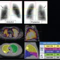
Dementia is a chronic, progressive neurodegenerative condition that affects multiple higher cognitive functional domains. There are several subtypes of dementia based on underlying pathology. Single-photon emission computed tomography (SPECT) demonstrates hypoperfusion patterns that differ among various dementia subtypes. These patterns are often analogous to patterns of hypometabolism seen on fluorodeoxyglucose positron emission tomography (FDG-PET).
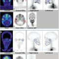
3.2.1 Alzheimer’s Disease
Posterior temporal and inferior parietal hypoperfusion generally precedes the onset of clinical symptoms (► Fig. 3.1 a). Precuneus hypoperfusion may be seen in some cases. A review of sagittal images is often helpful in diagnosis (► Fig. 3.1 b). Frontal hypoperfusion may be seen in advanced stages. There is usual sparing of the sensorimotor cortex and occipital lobes. Hypoperfusion may be asymmetric in the early stages and may extend to involve both hemispheres. 1

3.2.2 Frontotemporal Dementia
Bilateral frontal and anterior temporal hypoperfusion is seen in patients with predominantly behavioral variant frontotemporal dementia (► Fig. 3.2). Frontal hypoperfusion may also be seen in depression and schizophrenia, and clinical correlation is necessary to interpret SPECT findings.

3.2.3 Vascular Dementia
Patchy areas of hypoperfusion involving the cortical hemispheres or subcortical structures may be seen in patients with vascular dementia (► Fig. 3.3).

3.2.4 Primary Progressive Aphasia
Left temporal hypoperfusion (particularly in right-handed individuals) is seen in the semantic dementia variant of primary progressive aphasia (► Fig. 3.4). Semantic dementia is clinically characterized by a fluent aphasia and is associated with frontotemporal dementia-type pathology. However, patients with a logopenic variant of primary progressive aphasia (clinically characterized by slow speech, word retrieval difficulty, and speech paucity and dysfluency) may show an asymmetric Alzheimer’sdisease pattern, often affecting the language-dominant left hemisphere (► Fig. 3.5).


3.2.5 Corticobasal Degeneration and Progressive Supranuclear Palsy
In addition to cerebral cortex involvement, these syndromes may present with hypoperfusion in the basal ganglia involving the caudate and putamen.
3.2.6 Diffuse Lewy Body Disease
The hypoperfusion pattern in Lewy body dementia is similar to that seen in Alzheimer’s disease, except that there is prominent involvement of the occipital lobes.
Related posts:
Stay updated, free articles. Join our Telegram channel

Full access? Get Clinical Tree


