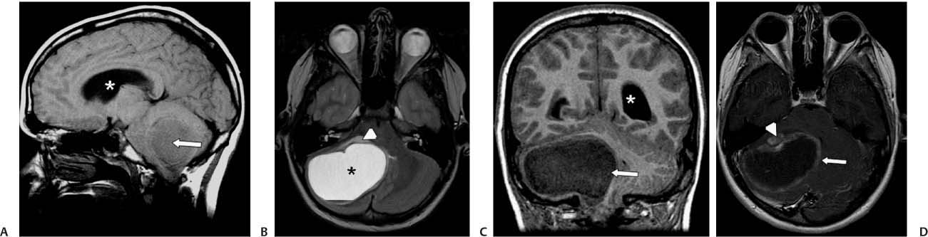
Case 30 A 13-year-old boy with a history of gait disturbance and vomiting. (A) Sagittal T1-weighted image (WI) shows an enlarged 4th ventricle. A mass (arrow) is effacing the 4th ventricle. The lateral ventricles are dilated (asterisk). (B) Axial T2WI shows a cystic mass centered on the right cerebellar hemisphere (asterisk). There is mild surrounding edema (arrowhead).(C) Coronal T1WI shows the right cerebellar mass (arrow) and the dilated lateral ventricle (asterisk). (D) Axial T1WI with contrast shows peripheral enhancement of the mass (arrow); there is an anterior mural nodule (arrowhead). • Pilocytic astrocytoma:
Clinical Presentation
Imaging Findings
Differential Diagnosis
![]()
Stay updated, free articles. Join our Telegram channel

Full access? Get Clinical Tree


