 Clinical Presentation
A 59-year-old miner with progressive dyspnea on exertion.
 Imaging Findings
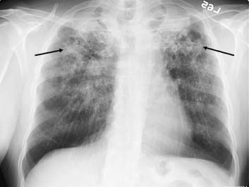
Conventional chest radiograph reveals extensive areas of confluent and masslike fibronodular opacities in both upper lobes, with calcification and apical pleural thickening (arrows). There is upper lobe volume loss bilaterally, with cephalic retraction of the pulmonary hila. An area of lucency is seen in the left upper lobe.
 Differential Diagnosis
• Silicosis: Complicated silicosis (progressive massive fibrosis) is characterized by masslike opacities resulting from the coalescence of fibronodular fibrosis. It tends to affect the upper lobes more often.
• Tuberculosis (TB): Bilateral apical fibronodular opacities with apical pleural thickening and upper lobe volume loss in particular are associated with cavitation and may be sequelae of chronic reactivation TB or atypical mycobacterial infection.
Stay updated, free articles. Join our Telegram channel

Full access? Get Clinical Tree


