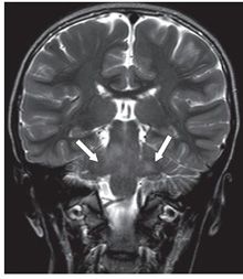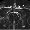
FINDINGS Figures 30-1 and 2. Axial and coronal T2WI through the brachium pontis, respectively. There is hyperintensity in the bilateral middle cerebellar peduncles (brachium pontis) (arrows). The pons is also significantly abnormal as well.
DIFFERENTIAL DIAGNOSIS Demyelinating disease, neurodegenerative disease, viral encephalitis, tumor, small vessel ischemia, phakomatosis.
DIAGNOSIS Phakomatosis (neurofibromatosis type 1).
DISCUSSION
Stay updated, free articles. Join our Telegram channel

Full access? Get Clinical Tree


