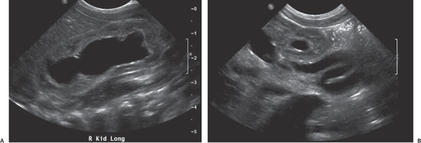
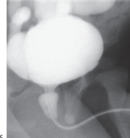
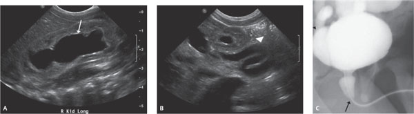
Case 30 A newborn with in utero hydronephrosis. (A,B) Sonographic images demonstrate severe hydronephrosis (arrow) and hydro-ureter (arrowhead). The findings were present bilaterally. (C) Oblique image from a voiding cysto-urethrogram demonstrates a bullet-shaped dilatation of the posterior urethra (arrow) with reflux into the (arrowhead) dilated ureter. The more distal urethra is of normal caliber. • Posterior urethral valves:
Clinical Presentation
Further Work-up
Imaging Findings
Differential Diagnosis
![]()
Stay updated, free articles. Join our Telegram channel

Full access? Get Clinical Tree


