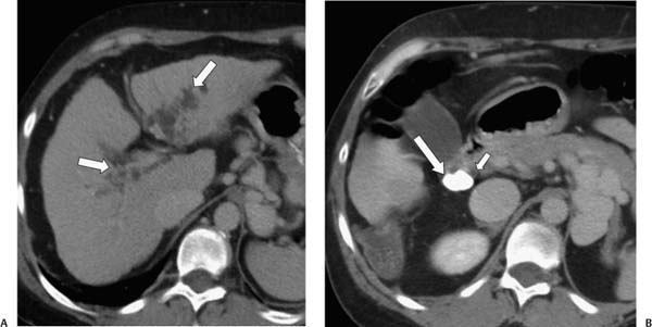
Case 30 A 47-year-old woman with a history of cirrhosis presents with jaundice, acute right upper quadrant abdominal pain, and fever. (A) Contrast-enhanced computed tomography shows bilateral intrahepatic biliary dilatation (arrows) and a shrunken liver, consistent with the provided history of cirrhosis. (B) A more caudal image shows a calcified stone (large arrow) within the expected location of the gallbladder neck or cystic duct. This stone abuts the common bile duct (CBD; small arrow). The incidental finding of a hypodense lesion in the visualized portion of the right lobe of the liver was determined to be hepatoma. •
 Clinical Presentation
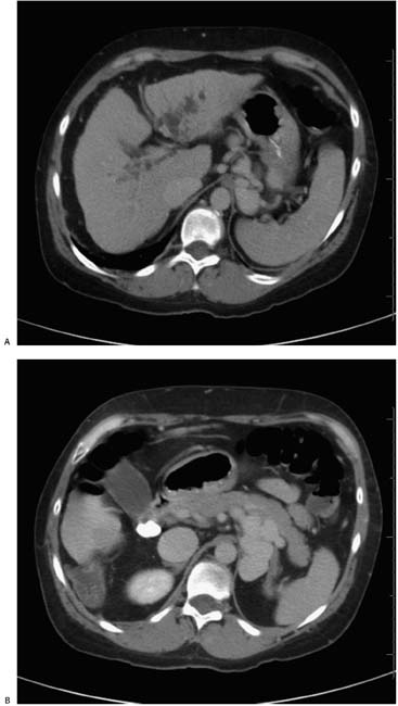
 Imaging Findings
 Differential Diagnosis
![]()
Stay updated, free articles. Join our Telegram channel

Full access? Get Clinical Tree


