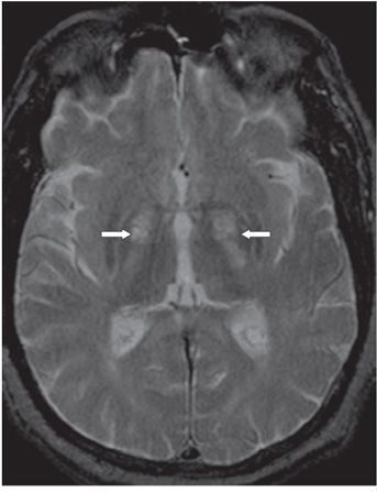
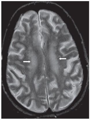
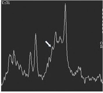
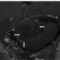
FINDINGS Figure 300-1. Axial T1WI through the basal ganglia. There is symmetrical hyperintensity in the globi pallidus (arrows). Figure 300-2. Axial T2WI in a different patient. There is hyperintensity in the medial globi pallidi (arrows). Figure 300-3. Axial T2WI through the centrum semiovale. There are patchy and nonspecific white matter abnormalities in both hemispheres (arrows). Figure 300-4. Short TE (31 ms) proton MR spectra show elevation of glutamine and glutamate (arrow).
DIFFERENTIAL DIAGNOSIS Hypoxic-ischemic encephalopathy, neurofibromatosis type 1, Japanese encephalitis, hyperalimentation or total parenteral nutrition (manganese deposition), hemorrhage (methemoglobin), Wilson disease (copper toxicity and subsequent gliosis), basal ganglia calcification, nonketotic hyperglycemia associated with chorea-balism, hypothyroidism, and carbon monoxide intoxication.
DIAGNOSIS Chronic hepatic encephalopathy (HE).
DISCUSSION


Stay updated, free articles. Join our Telegram channel

Full access? Get Clinical Tree


