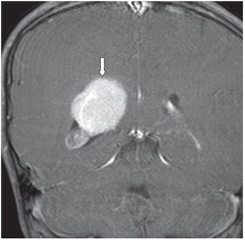
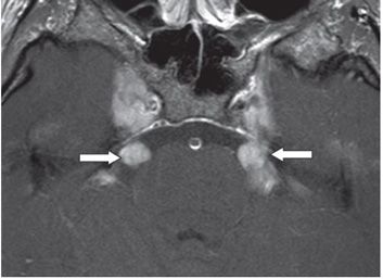
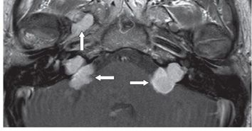
FINDINGS Figure 302-1. Coronal T2WI through the trigones. There is a 3.9-cm right trigonal isointense (to gray matter [GM]) mass (arrow) with surrounding peritrigonal vasogenic edema superiorly. Figure 302-2. Coronal post-contrast T1WI. The intraventricular mass enhances homogeneously and avidly. This is a surgically proven intraventricular meningioma. Figure 302-3. Axial post-contrast T1WI through the cavernous sinuses. There are bilateral lobulated contrast-enhancing masses along the path of the bilateral trigeminal nerves (transverse arrows). Figure 302-4. Axial post-contrast T1WI through the internal auditory canal (IAC). There are lobulated contrast-enhancing masses in bilateral cerebellopontine angle (CPA) extending into the IACs (transverse arrows). There is a similar contrast-enhancing smooth marginated mass in the region of the right foramen ovale (vertical arrow). Tumors in Figures 302-3 and 302-4 are presumed schwannomas of the trigeminal and vestibular nerves, respectively. Not shown here are right facial nerve and extensive schwannomas of the spinal nerves.
DIFFERENTIAL DIAGNOSIS Neurofibromatosis type 2 (NF2), Neurofibromatosis type 1 (NF1), schwannomatosis.


Stay updated, free articles. Join our Telegram channel

Full access? Get Clinical Tree


