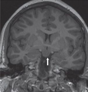
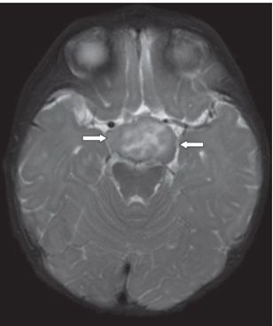
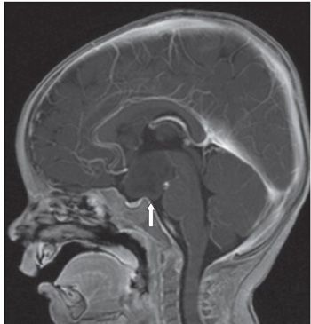
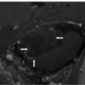
FINDINGS Figure 303-1. Sagittal non-contrast T1WI. There is a broad-based isointense mass (arrow) in the region of the tuber cinereum. Figure 303-2. Coronal non-contrast T1WI in the same patient shows that the mass has exactly the same signal as the gray matter (GM) (arrow). Figure 303-3. Axial T2WI in a companion case. There is a mass within the suprasellar cistern of heterogeneous intensity containing some central hyperintensities (arrows). Figure 303-4. Sagittal post-contrast T1WI shows the extent of the nonenhancing mass (arrow).
DIFFERENTIAL DIAGNOSIS Hypothalamic-chiasmatic glioma, craniopharyngioma, germinoma, and Langerhans cell histiocytosis, hamartoma of tuber cinereum.
DIAGNOSIS Hamartoma of tuber cinereum (HTC).
DISCUSSION


Stay updated, free articles. Join our Telegram channel

Full access? Get Clinical Tree


