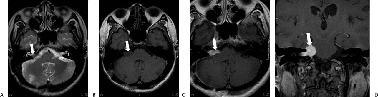
Case 31 A 43-year-old man with right-sided progressive hearing loss. (A) Axial T2-weighted image (WI) shows expansion of the right internal auditory canal (IAC). A mass is effacing the cerebrospinal fluid (CSF) inside the canal (arrow). (B) Axial T1WI without contrast shows a mass with intensity similar to that of the brain (arrow) in the right IAC and cerebellopontine angle(CPA). (C) Axial T1WI with contrast shows intense enhancement of the mass (arrow). (D) Coronal T1WI with contrast shows the enhancing mass in the right CPA and IAC (arrow). • Vestibular schwannoma:
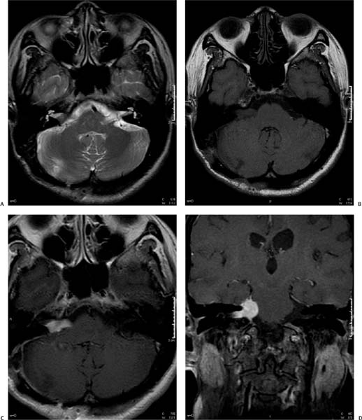
Clinical Presentation
Imaging Findings
Differential Diagnosis
![]()
Stay updated, free articles. Join our Telegram channel

Full access? Get Clinical Tree


