 Clinical Presentation
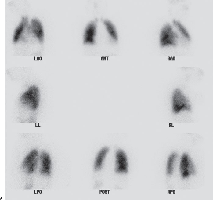
A 62-year-old woman with acute shortness of breath.
Further Work-up
Given the extrapulmonary activity, an additional spot image of the head was obtained to assess for intracranial activity.
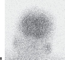

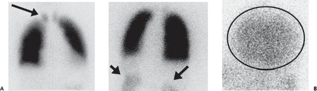
(A) Multiple Tc99m-MAA perfusion images demonstrate no moderate or large perfusion defects but show abnormal extrapulmonary activity in the thyroid and kidneys (arrows). (B) Spot image of the head demonstrates MAA accumulation in the cerebral cortex (circle).
 Differential Diagnosis
All three demonstrate extrapulmonary activity in the kidneys and thyroid. However, only a shunt will demonstrate uptake in the brain.
• Right-to-left shunt: Extrapulmonary uptake including kidneys, thyroid, and most importantly brain indicates a right-to-left shunt.
• Free pertechnetate:


Stay updated, free articles. Join our Telegram channel

Full access? Get Clinical Tree


