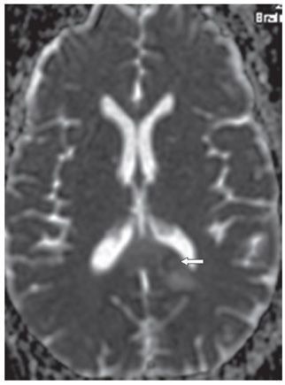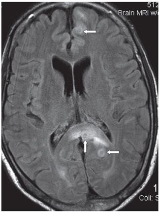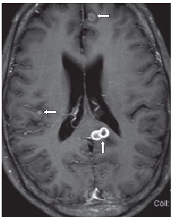


FINDINGS Figures 31-1 and 31-2. Axial DWI with corresponding ADC map. There is ring-restricted diffusion in the splenium of the corpus callosum (CC) (arrows). There is surrounding increased diffusion consistent with vasogenic edema. Figure 31-3. Axial FLAIR through the splenium of the CC. There is smudgy hyperintensity through the splenium of the CC surrounding a hyperintense ring with hypointense core (vertical arrow). There is an additional ring hyperintensity in the adjacent left occipital lobe with a hypointense core (posterior transverse arrow) and in the parafalcine left frontal lobe (anterior transverse arrow). Figure 31-4. Axial post-contrast T1WI through the splenium just superior to Figure 31-3. There are two adjacent ring contrast enhancing lesions in the splenium on the left with hypo/isointense core (arrow) and other ring-enhancing cortical lesions elsewhere in the brain (transverse arrows). These also have isointense core.
DIFFERENTIAL DIAGNOSIS Tuberculomas (TBs), metastasis, pyogenic abscess, fungal infection, neurocysticercosis.
DIAGNOSIS CC TB.
Stay updated, free articles. Join our Telegram channel

Full access? Get Clinical Tree


