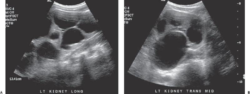
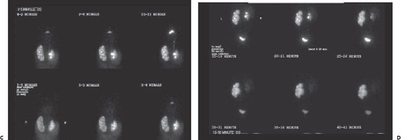
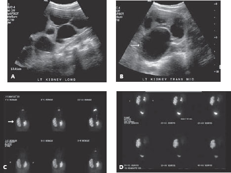
Case 31 An infant with hydronephrosis on prenatal ultrasound. (A,B) Sonographic images of the left kidney demonstrate multiple ovoid, anechoic structures. Note the dilated central anechoic structure, consistent with a dilated renal pelvis (arrow), making this more likely to be hydronephrosis than multiple cysts. (C,D) Images from a MAG3 (mercapto-acetyl-triglycine) nuclear medicine study demonstrate the delayed uptake and excretion of radioactive tracer by the enlarged left kidney. Note the dilated calyces (arrow). • Ureteropelvic junction (UPJ) obstruction:
Clinical Presentation
Further Work-up
Imaging Findings
Differential Diagnosis
![]()
Stay updated, free articles. Join our Telegram channel

Full access? Get Clinical Tree


