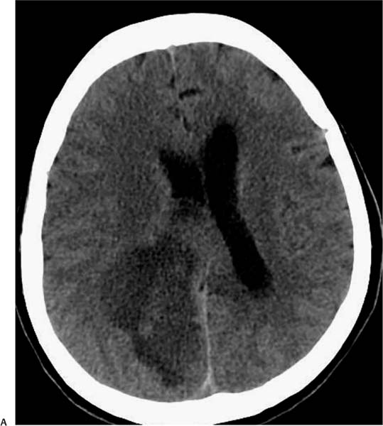
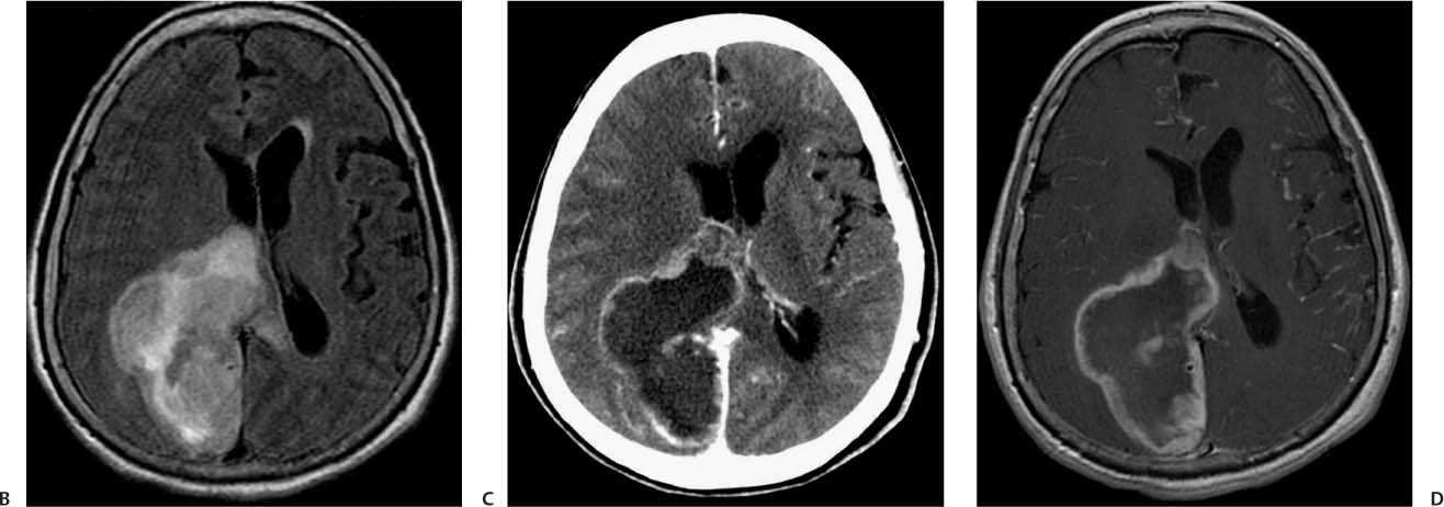
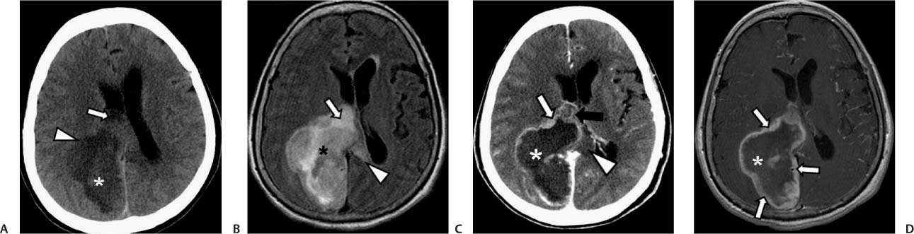
Case 32 A 64-year-old woman with progressive mental deterioration, nausea, and vomiting. (A) Axial computed tomography (CT) scan of the head without contrast shows a low-density mass in the right occipital region (asterisk) with an isodense halo (arrowhead) and mass effect that effaces the lateral ventricle (arrow). (B) Axial fluid-attenuated inversion recovery (FLAIR) image shows a heterogeneous mass (asterisk) with peripheral high signal that crosses the corpus callosum (arrowhead). The mass effaces the lateral ventricle (arrow). (C) Axial CT scan of the head with contrast shows irregular, thick peripheral enhancement (white arrow) and the low-density mass (asterisk). The edema crossing the corpus callosum is seen (arrowhead). The mass has subependymal extension to the lateral ventricle (black arrow). (D) Axial T1-weighted image (WI) with contrast shows irregular enhancement (arrows) of the right occipital mass (asterisk). • Glioblastoma multiforme (GBM):
Clinical Presentation
Further Work-up
Imaging Findings
Differential Diagnosis
![]()
Stay updated, free articles. Join our Telegram channel

Full access? Get Clinical Tree


