 Clinical Presentation
A 34-year-old man with chronic cough.
 Imaging Findings
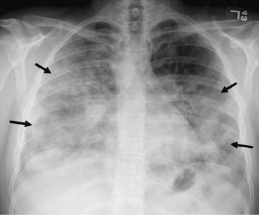
Chest radiograph shows dense, multifocal parenchymal ground-glass opacities and denser areas of consolidation that are more confluent in the mid and lower lung zones. There is relative sparing of the apices (arrows). There is no evidence of pleural fluid, and the cardiac silhouette is normal in size. Incidental note is made of a right-sided aortic arch.
 Differential Diagnosis
• Pulmonary alveolar proteinosis (PAP): Ground-glass opacity with air-space consolidation in a bilateral and multifocal distribution is a common imaging presentation of PAP.
• Pulmonary edema:
Stay updated, free articles. Join our Telegram channel

Full access? Get Clinical Tree


