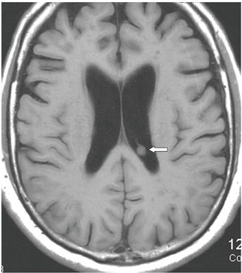
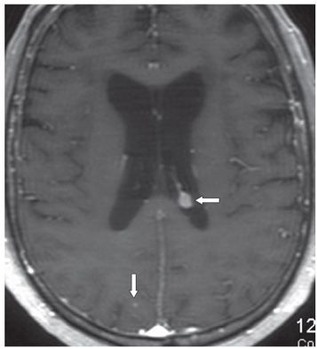
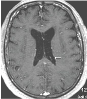
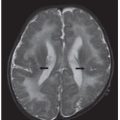
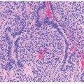
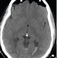
FINDINGS Figures 32-1 to 32-3. Axial T2WI, non-contrast T1WI, and post-contrast T1WI through the lateral ventricles, respectively. There is a small left lateral ventricle round T1 and T2 isointense (to white matter [WM]) avidly contrast-enhancing choroid plexus mass (arrows). There is also a punctate metastasis in the right parasagittal parietal lobe (vertical arrow in Figure 32-3). Numerous new punctate contrast-enhancing metastases are present in other areas of the brain (not shown). Figure 32-4. Axial post-contrast T1WI through same level 7 months before. There is no significant mass in the choroid plexus.
DIFFERENTIAL DIAGNOSIS Intraventricular meningioma, colloid cyst, xanthogranuloma, choroid plexus metastasis.
DIAGNOSIS Metastasis non-small cell lung carcinoma.


Stay updated, free articles. Join our Telegram channel

Full access? Get Clinical Tree


