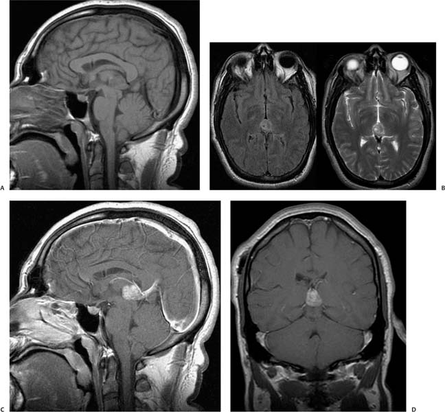
Case 33 A patient with precocious puberty and limited upward gaze. (A) Sagittal T1-weighted image (WI) shows a mass in the pineal region (arrow) compressing the tectum (arrowhead). There is a ventricular shunt in place to drain hydrocephalus from aqueductal compression. (B) Axial T2WI shows a heterogeneous mass in the pineal region (arrow). The mass has punctate areas of hypointensity (calcifications). (C) Sagittal T1WI with contrast shows diffuse enhancement of the mass (arrow). (D) Coronal T1WI with contrast shows the enhancing mass in the pineal region (arrow). • Germinoma:
Clinical Presentation
Imaging Findings
Differential Diagnosis
![]()
Stay updated, free articles. Join our Telegram channel

Full access? Get Clinical Tree


