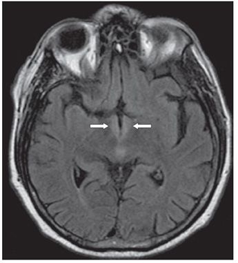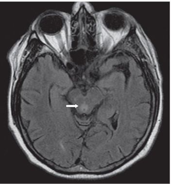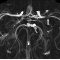

FINDINGS Figure 33-1. Axial FLAIR through the third ventricle. There is bilateral symmetrical hyperintensity (arrows) along the walls of the third ventricle. Figure 33-2. Axial FLAIR slightly below Figure 33-1. There is hyperintensity in the hypothalamus (arrows) and dorsal midbrain. Figure 33-3. Axial FLAIR through the midbrain. There is periaqueductal hyperintensity (arrow).
DIFFERENTIAL DIAGNOSIS Top of the basilar syndrome, deep venous system thrombosis, neuromyelitis optica, viral encephalitis, Wernicke encephalopathy (WE), acute disseminated encephalomyelitis, Creutzfeldt-Jacob disease, mitochondrial disorder, and toxic-related changes (metronidazole).
Stay updated, free articles. Join our Telegram channel

Full access? Get Clinical Tree


