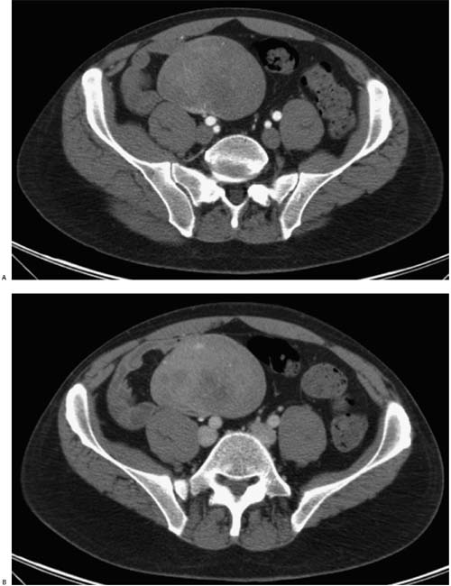
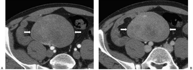
Case 33 A 52-year-old man with an abdominal mass palpated during a routine physical examination. (A) Arterial-phase contrast-enhanced computed tomography (CT) shows a large, well-circumscribed, homogeneous mass (arrows) in the pelvic mesentery. (B) Venous-phase image shows heterogeneous enhancement of this mass (arrows). • Gastrointestinal stromal tumor (GIST): GIST is the first choice for a smoothly marginated mass in the mesentery with heterogeneous enhancement. • Desmoid tumor (mesenteric fibromatosis):
 Clinical Presentation
 Imaging Findings
 Differential Diagnosis
![]()
Stay updated, free articles. Join our Telegram channel

Full access? Get Clinical Tree


