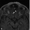
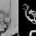
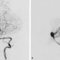
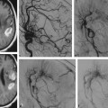
Fig. 33.1 MRA (a) reveals a left cavernous dAVF, draining into the superior ophthalmic vein, further draining into the angular vein and common facial vein, respectively. No cortical venous reflux is identified. Left external carotid artery (ECA) (b) and left ICA (c) angiograms in lateral view show supply to the dAVF from the artery of the foramen rotundum, petrous branches of the middle meningeal artery, and the meningohypophyseal trunk of the ICA. Treatment was deemed necessary in this case, despite the benign type, because of the increased ocular pressure.
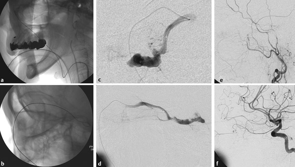
Fig. 33.2 Skull radiography in lateral views (a,b) demonstrates the approach from the internal jugular vein through the common facial vein into the superior ophthalmic vein to reach the cavernous sinus. Microcatheter injections in anteroposterior (AP) (c) and lateral (d) views confirm the location of the microcatheter within the cavernous sinus. Left ECA (e) and left ICA (f) angiograms in lateral view after coil embolization demonstrate complete obliteration of the cavernous dAVF.
33.1.3 Diagnosis
Left cavernous dural arteriovenous fistula (dAVF).
33.2 Embryology and Anatomy
The cavernous sinus can be defined at around the 18-mm stage, arising from the anterior or trigeminal portion of the primitive head vein. At the 20-mm stage, it receives tributaries from the ophthalmic veins and a large cerebral vein draining the lateral wall of the diencephalon, as well as smaller tributaries from the network in the region of the semilunar ganglion. At this point, no tributaries are detected from the cerebral hemisphere, as the blood now flows in the opposite direction, toward the developing transverse sinus. The interruption between the cavernous sinus and the internal jugular vein (the trunk of the primitive head vein) is also complete, and the blood from the cavernous sinus flows upward through the petrosquamous sinus (remnant of the trunk of the middle dural plexus that will usually regress or may persist as a connection between the cavernous sinus and the middle meningeal vein) into the transverse sinus.
The superior petrosal sinus initially forms as a tributary of the petrosquamous sinus and only later connects the cavernous sinus with the transverse sinus. A small plexiform inferior petrosal sinus can be seen at around the 14-mm stage, surrounding cranial nerves IX and X and becoming an apparent drainage pathway connecting the cavernous sinus to the internal jugular vein at around the 50-mm stage.
In the fetus, at around the 70–128-mm stages (13–18 weeks’ gestation), the cavernous sinus is a complex network consisting of many fine tubular venous spaces (also known as the venous canals of Krivosic), which develop in the complex immature lateral sella mesenchyme. These spaces are formed only by an endothelial layer, with no smooth muscles, unlike the common venous walls. As the development progresses to around the 180-mm stage (23 weeks’ gestation), these venous channels expand with progressive thinning of the immature mesenchyme, which is taken over by the development of the dura and collagen after the stage of 230 mm (28 weeks’ gestation). Depending on the variations in further development, the cavernous sinus in an adult may therefore be a large sinusoidal venous space containing trabeculations or a venous plexus consisting of multiple venous channels.
Inferior draining pathways from the cavernous sinus connecting to the pterygoid plexus have been observed from the 70-mm stage (13 weeks’ gestation). The number of these venous pathways abruptly increases after the 230-mm stage (28 weeks’ gestation), likely related to the rapidly developing skull base and its neural foramina.
Two secondary intradural anastomoses involving the cavernous sinus usually occur postnatal or in the late fetal phase. The first one is between the embryonic tentorial sinus (draining the middle cerebral veins) and the anterior end of the cavernous sinus, near its junction with the common ophthalmic vein. This anastomosis will eventually link the superficial and deep middle cerebral veins (i.e., the cerebral hemispheric drainage) with the cavernous sinus and, therefore, with the basal vein, respectively. This is also known as the “cavernous capture,” which typically occurs during the first year of life. The second anastomosis, commonly dorsal to the fifth nerve root, is between the cavernous sinus and the superior petrosal sinus at the entrance of the great anterior cerebellar vein (or metencephalic vein).
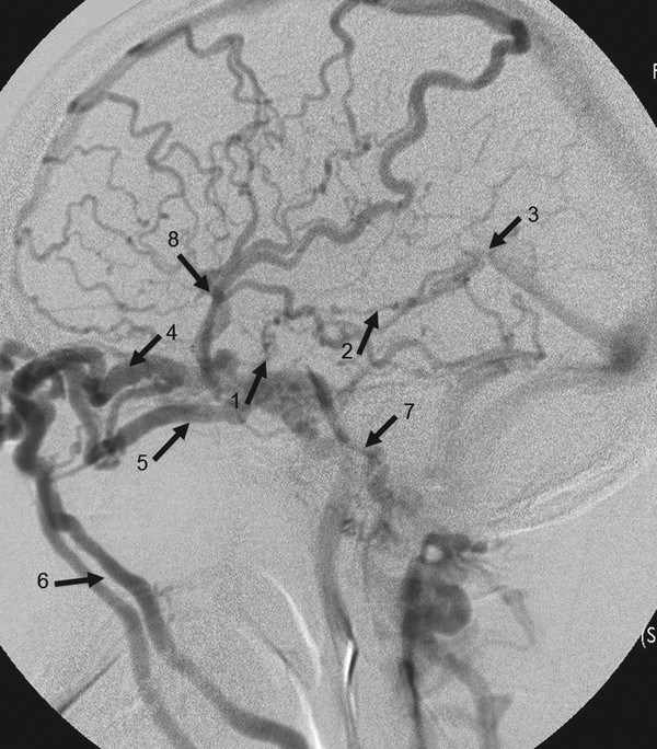
In an adult, the cavernous sinus typically has the following connections (▶ Fig. 33.3; ▶ Fig. 33.4): First, superoanterior with the ophthalmic veins, middle cerebral vein, or superficial Sylvian vein, and from the same superior region but directed posteriorly, via the deep middle cerebral vein/uncal vein to the basal vein of Rosenthal; second, inferiorly with the pterygoid plexus through various neural foramina at the skull base, the most prominent of which being the vein of the foramen ovale; and third, posteriorly with the superior petrosal vein, which may connect with the petrosal vein of the posterior fossa; the inferior petrosal vein, which connects with the internal jugular vein; and the basilar plexus, which connects with the vertebral venous plexus. Finally, it connects medially with the contralateral cavernous sinus through at least two intercavernous anastomotic channels.
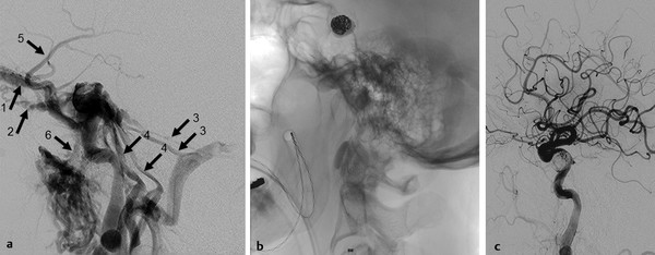
Fig. 33.3 ICA injection in the lateral view (a) of a traumatic carotid-cavernous fistula demonstrates in this patient the following routes of drainage of the cavernous sinus: anteriorly, there is drainage through the superior and inferior ophthalmic veins (1,2), and posteriorly and inferiorly, there is drainage to the bilateral superior (3) and inferior (4) petrous sinuses to the sigmoid sinus and jugular vein. Laterally and superiorly, there is drainage toward the superficial middle cerebral vein (5) to the cortical veins, and anteroinferiorly, there is drainage via the vein of the foramen ovale (6) to the pterygoid plexus. After a combination of coil and balloons (b), the fistula could be occluded (c).
