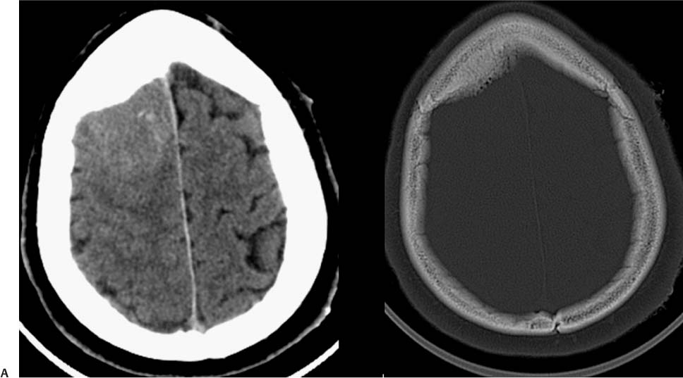
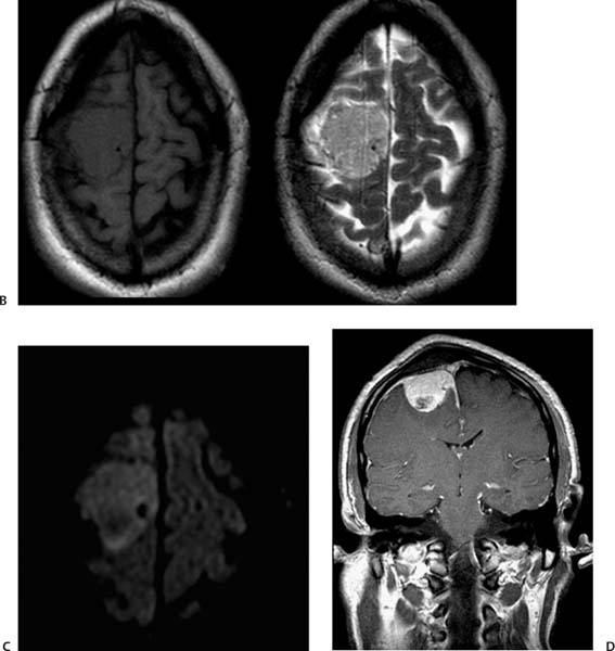
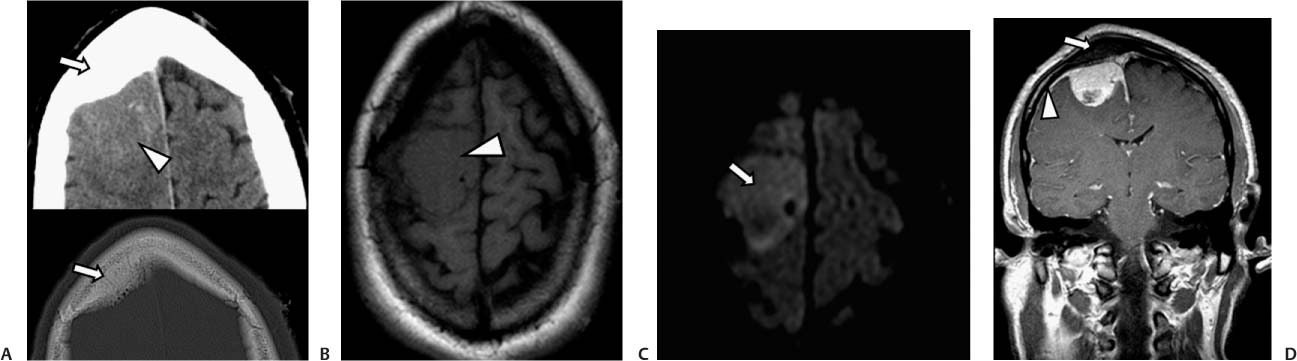
Case 34 A 48-year-old man with left-sided motor weakness and vomiting. (A) Axial computed tomography (CT) scan of the head shows a slightly hyperdense lesion in the right frontal region (arrowhead) with adjacent cortical thickening (arrows). (B) Axial T1-weighted image (WI) shows an isointense lesion in the right frontal region (arrowhead) that effaces the adjacent sulci. (C) Diffusion-WI shows increased signal in the right frontal lesion (arrow). (D) Coronal T1WI with contrast shows diffuse enhancement of the mass. There is thickening of the adjacent skull (arrow). The arrowhead shows the dural tail. • Meningioma:
Clinical Presentation
Further Work-up
Imaging Findings
Differential Diagnosis
![]()
Stay updated, free articles. Join our Telegram channel

Full access? Get Clinical Tree


