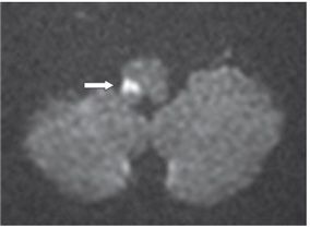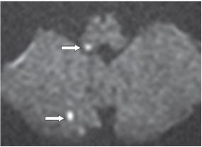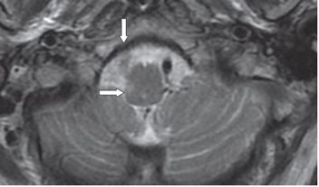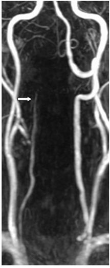


FINDINGS Figures 34-1 and 34-2. Axial DWI through the medulla. There is a right posterolateral medulla small area of restricted diffusion (arrows). Figure 34-3. Axial DWI through the restiform body. There is focal restricted diffusion in the right restiform body (anterior arrow) with punctate areas of restricted diffusion in the right cerebellum (posterior arrow). Figure 34-4. Axial T2WI through the medulla. There is a small right lateral medullary hyperintensity (transverse arrow). Signal void is missing within the right vertebral artery (vertical arrow). Figure 34-5. Contrast-enhanced MRA of the neck. There is tapered occlusion of the right distal vertebral artery (V2) consistent with dissection.
DIFFERENTIAL DIAGNOSIS
Stay updated, free articles. Join our Telegram channel

Full access? Get Clinical Tree


