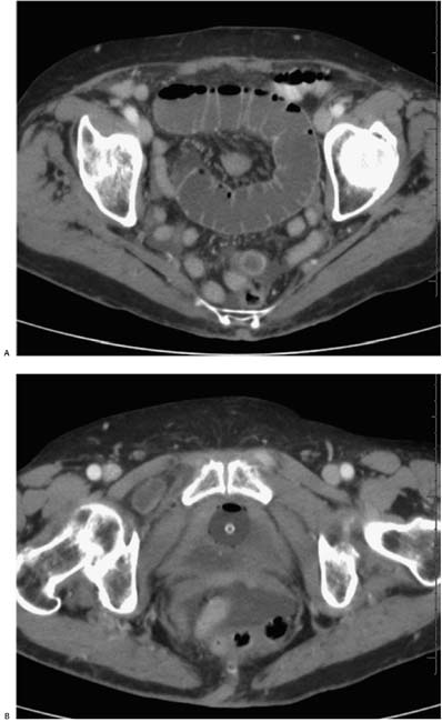
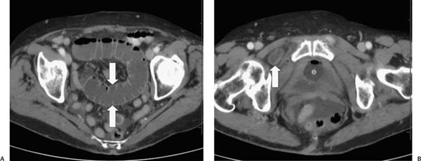
Case 34 A 91-year-old woman presents to the emergency department with intense abdominal pain. (A) Contrast-enhanced computed tomography (CT) shows a markedly dilated loop of small bowel in the pelvis (arrows). (B) More caudal slice shows a segment of small bowel (arrow) between the external obturator and pectineus muscles. • Obturator foramen hernia: This is the only diagnosis based on the finding of small-bowel obstruction, dilated small bowel, as well as a fluid-filled structure in the obturator foramen. • Pelvic external hernias are congenital, traumatic, or post-surgical herniations of pelvic contents through the pelvic wall or groin. They include inguinal, femoral, obturator, sciatic, and perineal hernias.
 Clinical Presentation
 Imaging Findings
 Differential Diagnosis
 Essential Facts
Stay updated, free articles. Join our Telegram channel

Full access? Get Clinical Tree


