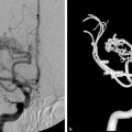
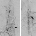
Fig. 34.1 Right internal carotid artery (ICA) angiogram in lateral view in arterial (a) and late capillary (b) phases show a frontal AVM nidus with drains cranially to the superior sagittal sinus via the cranial segment of the vein of Trolard. Because of cranial venous outflow obstruction, the AVM also drains through the caudal segment of the vein of Trolard that connects to the superficial middle cerebral vein (arrow). This anastomosis allows the nidus to drain finally to the vein of Rosenthal (arrowheads) via the deep middle cerebral vein (thin double arrows).
34.1.3 Diagnosis
Right frontal AVM with codominant vein of Trolard and superficial middle cerebral vein (SMV).
34.2 Embryology and Anatomy
The superficial venous draining system is composed of a network of subpial veins, which run underneath the arterial network, opening into larger subarachnoid venous collectors (typically called cortical draining veins) and finally drain into the dural venous sinuses. These veins drain the brain cortex and superficial white matter, including the U-fibers.
Anatomic variations are the norm, rather than the exception, and cortical veins vary in size, number, route, and drainage pathway. Naming individual veins according to their topographic location is not practical and has led to many different names, for inconstant arrangements. From a practical approach, the superficial venous system can be subdivided into three functional groups that are in hemodynamic equilibrium with each other and that may be interconnected. Each of these groups has its own major venous collector, which can provide an alternative anastomotic pathway to the other groups.
First is the mediodorsal group: It collects the high-convexity and midline cortical veins, opening preferentially into the superior sagittal sinus. The major venous collector for this group is the vein of Trolard.
Second is the posteroinferior group, which collects the parietotemporal region and opens preferentially into the transverse sinus. The venous collector for this group is the vein of Labbé.
Third is the anterior group. It collects the veins along the sylvian fissure and opercular region. The venous collector for this group is the SMV.
The vein of Trolard, also called the superior anastomotic vein, is by definition the largest anastomotic venous channel connecting the SMV with the superior sagittal sinus. It usually lies over the precentral, central, or postcentral sulcus but may be as far anterior as the anterior frontal veins or as far posterior as the anterior parietal veins. A “duplicated” vein of Trolard may be present, in which case two equal-sized anastomotic veins connect the SMV territory with the superior sagittal sinus.
The vein of Labbé, also known as inferior anastomotic vein, is by definition the largest venous channel connecting the SMV with the transverse sinus. Classically, it will course over the occipitotemporal sulcus, along the path of the middle temporal vein. Less frequently, it will course along the path of the anterior or posterior temporal veins. A “duplicated” vein of Labbé may be present, but the posterior vein will typically be larger.
The SMV, also called the superficial sylvian vein, overlies the sylvian fissure and drains the region of the operculum. Most frequently, it drains anteriorly through the venous sinuses of the sphenoid ridge into the cavernous sinus or the pterygoid venous plexus. It also may drain directly into the cavernous sinus or course around the temporal pole to drain into the superior petrosal sinus, through the inferior temporal vein. Superiorly, it may anastomose with the vein of Trolard, and posteroinferiorly, with the vein of Labbé. Two separate trunks of the SMV may be seen, but these will classically converge into a single channel more anteriorly. The central segment of the SMV may be absent. In this case, the anterior component will drain toward the sphenoid ridge sinuses and the posterior segment to the vein of Trolard, the vein of Labbé, or both.
Stay updated, free articles. Join our Telegram channel

Full access? Get Clinical Tree


