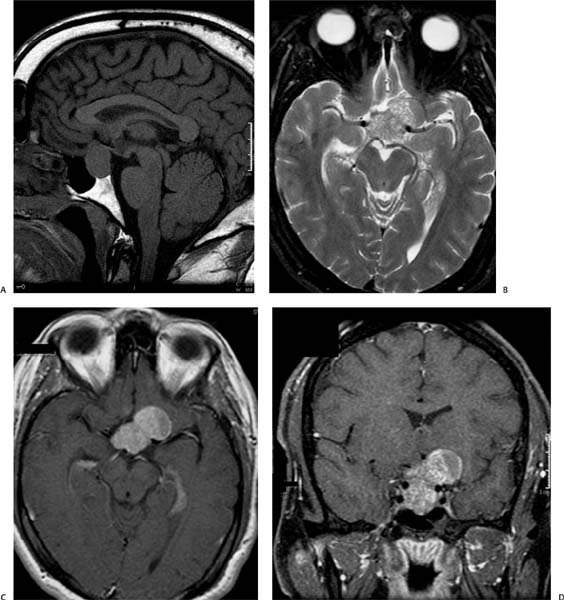
Case 35 A 43-year-old with headache and bitemporal hemianopsia. (A) Sagittal T1-weighted image (WI) shows a sellar mass with suprasellar extension (asterisk). The mass is isointense to brain and is displacing the optic chiasm superiorly (arrow). (B) Axial T2WI shows a heterogeneous mass in the suprasellar region (asterisk) that encases the internal carotid arteries without obstructing their flow (arrowheads). (C) Axial T1WI with contrast shows avid enhancement of the mass (asterisk). (D) Coronal T1WI with contrast show the encasement of the internal carotid arteries (arrowheads) by the mass (asterisk). • Pituitary macroadenoma:
Clinical Presentation
Imaging Findings
Differential Diagnosis
![]()
Stay updated, free articles. Join our Telegram channel

Full access? Get Clinical Tree


