 Clinical Presentation
A rising creatinine despite good urine output status post renal transplant.
Further Work-up
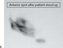

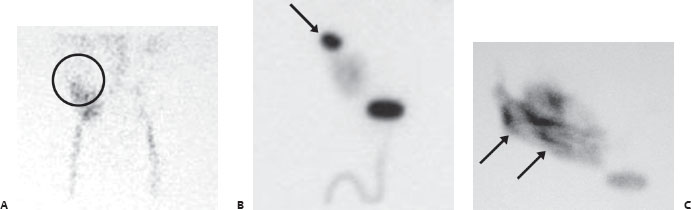
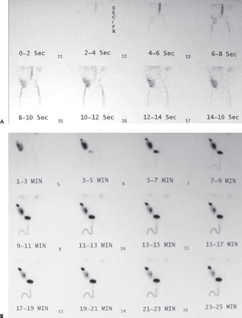
(A) Anterior pelvic angiographic phase images from a Tc99m MAG3 scan demonstrate prompt perfusion (within 2 seconds of iliac perfusion) to a renal transplant in the right iliac fossa. However, there is a lack of demonstrable flow to the upper pole. As a result, the superior portion ends with an unusual concave appearance (circle). (B) Function-phase images demonstrate normal uptake and excretion by the middle and inferior renal portions. However, there is progressive focal accumulation of radiolabeled urine at the superior portion (arrow). (C) Following patient repositioning, there is free spillage of the collection within the pelvic peritoneal cavity (arrows).
 Differential Diagnosis
• Upper pole infarct and partially contained urinary leak:


Stay updated, free articles. Join our Telegram channel

Full access? Get Clinical Tree


