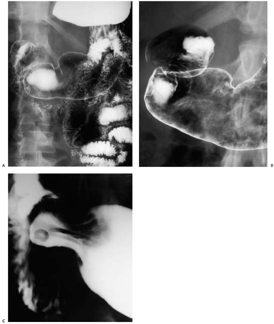
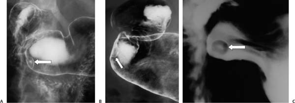
Case 35 A 40-year-old man presents for an upper gastrointestinal barium study to evaluate chronic, mild midepigastric pain. (A) Double-contrast barium study shows a submucosal lesion (arrow) in the gastric antrum, slightly in profile and slightly en face. A small collection of barium is seen within the center of the lesion. (B) Tangential view of the lesion (arrow) shows it to be focal and submucosal. (C) Single-contrast view en face shows the ovoid lesion with a central contrast collection (arrow). • Ectopic pancreatic rest: This is the most likely diagnosis. It is a classic non-neoplastic cause of a single bull’s-eye lesion. • Spindle cell or stromal tumor: These tumors can be singular, submucosal, and centrally necrotic/ulcerated, including leiomyoma and gastrointestinal stromal tumor (GIST). These neoplasms are typically larger on presentation. • Primary malignancy: These may be small, solitary, and ulcerated, such as adenocarcinoma, lymphoma, or carcinoid.
 Clinical Presentation
 Imaging Findings
 Differential Diagnosis
Stay updated, free articles. Join our Telegram channel

Full access? Get Clinical Tree


