The Transmedullary Veins
35.1 Case Description
35.1.1 Clinical Presentation
A 61-year-old previously healthy man came to medical attention because of mild cognitive deficits and slight subjective gait instability.
35.1.2 Radiological Studies
See ▶ Fig. 35.1, ▶ Fig. 35.2, and ▶ Fig. 35.3.
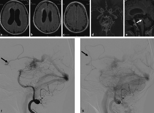
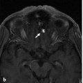
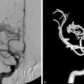
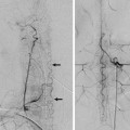
Fig. 35.1 Axial fluid-attenuated inversion recovery (FLAIR) weighted scans in multiple cuts (a,b,c) demonstrate mild hydrocephalus with increased size of the ventricles, decreased width of the sulci, and periventricular FLAIR hypersignal as a sign for decreased transependymal CSF reabsorption. The time-of-flight MRA (d) shows a choroidal AVM, and the midsagittal T1-weighted scan (e) proves patency of the aqueduct (arrow). No other cause for the patient’s hydrocephalus was identified. Conventional angiography (vertebral artery injection, lateral views on early and late phases f,g) revealed the AVM, which drained not only toward the straight sinus but also into the transmedullary veins (arrows in f,g). Case continued in ▶ Fig. 35.2.
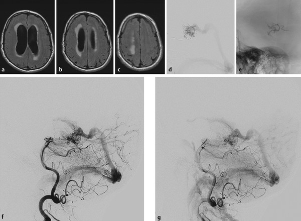
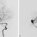
Fig. 35.2 Within the next 5 months, the patient’s symptoms became worse, with significant impairment of cognitive function and corresponding worsening of his hydrocephalus (axial FLAIR weighted scans, a,b,c), with ventricular widening and increased interstitial edema suggesting further decreased transependymal CSF reabsorption. There was no evidence for obstruction of the ventricular system, and it was proposed that the hyperpressure in his transependymal veins after arterialization was related to an impaired CSF reabsorption through the transmedullary veins. After partial embolization ([d] microcatheter position in the posterior lateral choroidal artery; [e] glue cast) that led to obliteration of the transmedullary drainage (conventional angiography postembolization, lateral view [f,g]), the symptoms reversed. Case is continued in ▶ Fig. 35.3.

Fig. 35.3 MR follow-up 3 weeks after the embolization demonstrated marked improvement of the transependymal CSF reabsorption on various axial FLAIR sections (a–d). The hydrocephalus had decreased, indicating it was indeed the arterialization of the transmedullary veins that could be held responsible for the decreased CSF reabsorption.
35.1.3 Diagnosis
Choroidal arteriovenous malformation (AVM) with reflux into transmedullary veins leading to hydrocephalus.
35.2 Embryology and Anatomy
The transcerebral veins are also called medullary or anastomotic veins, as they interconnect the superficial with the deep venous systems. Embryologically, these veins become apparent in the 40-mm embryo as a plexus of fine, straight veins that pass from the ependymal layer to the surface of the cortex, where they drain into pial collectors. There are two transcerebral venous systems present that may interconnect with each other via deep-seated venular anastomoses: the superficial and the deep medullary veins. While the former drain the white matter toward the surface from a depth of 1–2 cm, the latter drain the remainder of the white matter toward the ventricle centripetally into the subependymal veins of the lateral sinuses. Direct anastomotic veins between the superficial cortical and deep subependymal systems may be present.
The deep veins of the frontal and parietal lobes have a fan-shaped organization and converge at the superolateral angle of the lateral ventricle, with frontal veins joining the septal and anterior caudate veins and the parietal veins draining toward the thalamostriate veins. The occipital lobe veins join the lateral atrial veins, whereas the temporal lobe transmedullary veins take an ascending direction to join the inferior ventricular veins.
Stay updated, free articles. Join our Telegram channel

Full access? Get Clinical Tree


