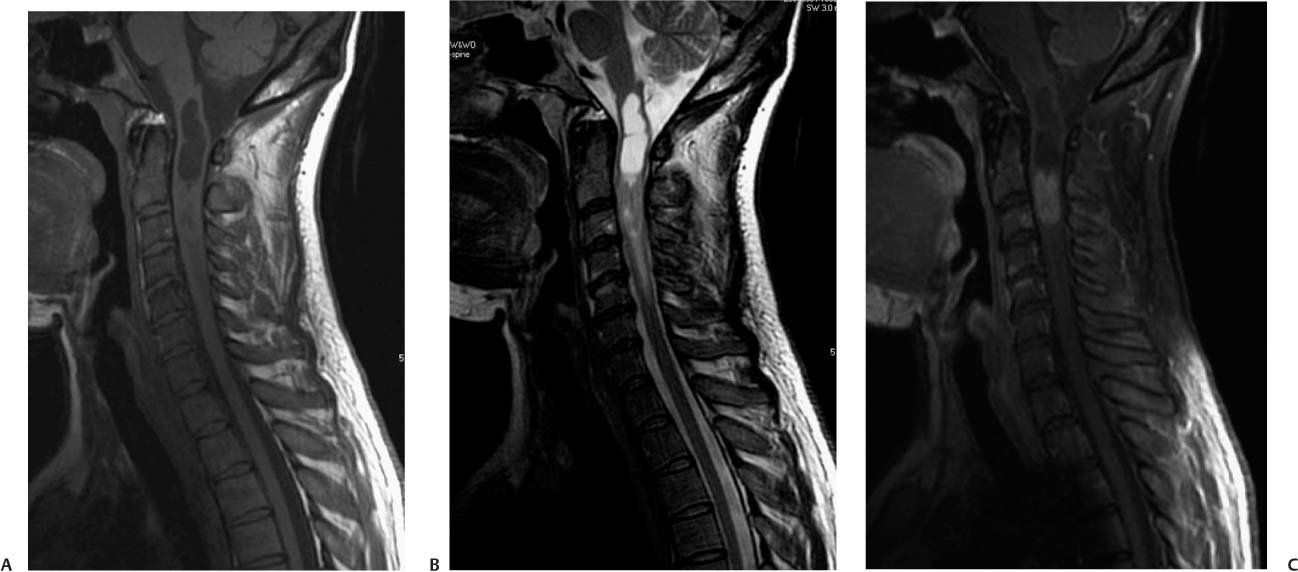
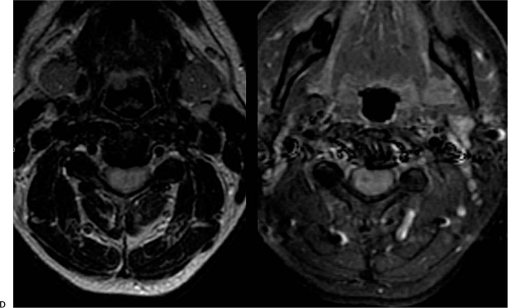
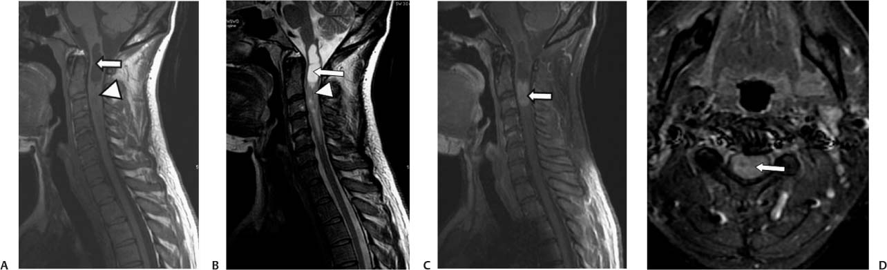
Case 36 A 38-year-old woman with progressive bilateral lower extremity paresthesia. D (A) Axial T1-weighted image (WI) of the cervical spine shows a syrinx (arrow) proximal to an ill-defined mass that thickens the spinal cord (arrowhead). (B) Sagittal T2WI of the cervical spine shows an ill-defined intramedullary mass (arrowhead) that is hyperintense to the spinal cord and proximal syringomyelia (arrow). (C) Sagittal T1WI of the cervical spine with contrast shows an enhancing mass at the C2-C3 level (arrow). (D) Axial T1WIs of the cervical spine with contrast, without and with fat suppression, show a thickened spinal cord with a diffuse intramedullary enhancing mass (arrow). • Intramedullary astrocytoma:
Clinical Presentation
Further Work-up
Imaging Findings
Differential Diagnosis
![]()
Stay updated, free articles. Join our Telegram channel

Full access? Get Clinical Tree


