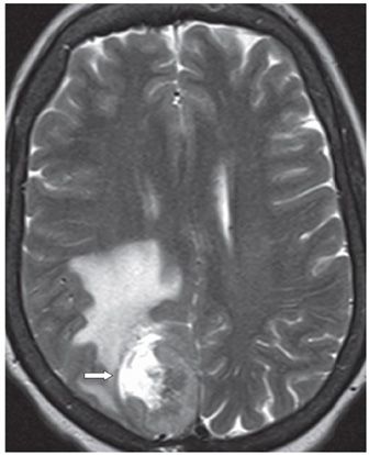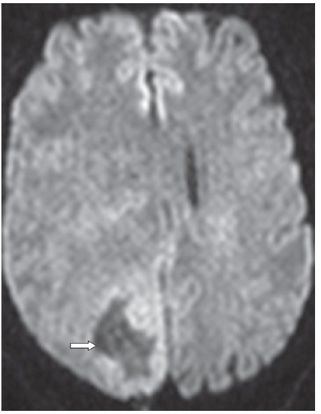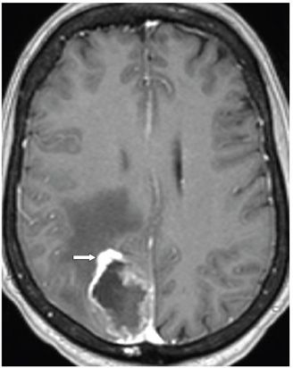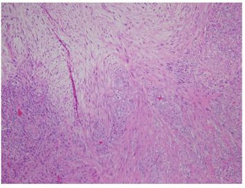


FINDINGS Figure 36-1. Axial T2 FLAIR. There is a right parasagittal parietal cortical mass with dural abutment. Confluent hyperintensity is present anteriorly and laterally to the mass most consistent with vasogenic edema or infiltrating tumor (vertical arrow). The mass is heterogeneous but mostly hyperintense with some inner rind of hypointensity (transverse arrow). Figure 36-2. Axial T2WI is very similar to the T2 FLAIR image. There is cerebrospinal fluid (CSF) hyperintensity anteriorly and laterally within the mass consistent with area of necrosis versus cyst formation (arrow). Figure 36-3. Axial DWI through the mass. There is mild hyperintensity in the medial and posterior rim of the mass that corresponds with minimally elevated to normal ADC values. The T2 hyperintensity anterior to the mass has markedly elevated ADC values (arrow). The findings correspond to a cystic necrotic mass with greater solid component posteriorly and medially with a rind of debris. Gradient echo image that is not shown was unremarkable for calcifications or blood product. Figure 36-4
Stay updated, free articles. Join our Telegram channel

Full access? Get Clinical Tree


